Osteoid Osteoma in the Front of the Mandible
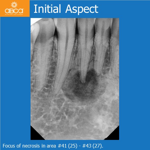
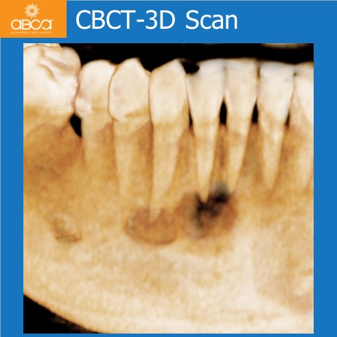
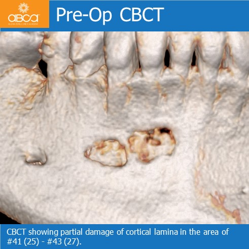
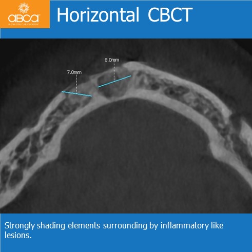
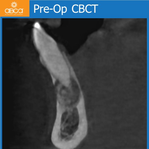
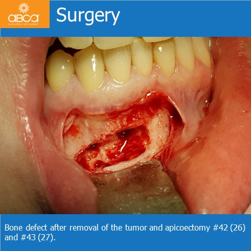
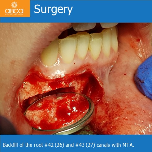
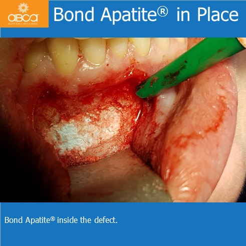
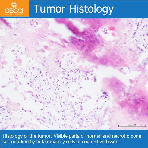
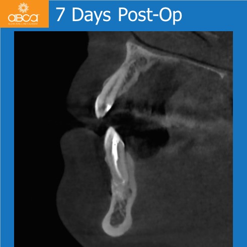
The patient is a 33 year old female, who complained of pain on the right side of the front mandible. The first symptom was pain similar to pulpitis in teeth #41 (25), #42 (26) and #43 (27). After endodontic treatment, the X-rays and CBCT showed a focus of necrotizes with strongly sheading elements in the central area. We did a root resection with backfill of the root canal with MTA in teeth #42 (26) and #43 (27). The tumor was removed with peripheral bone curettage to about 1 millimeter deep. The biological structure of the tumor was similar to spongy bone, but little harder. The bone defect was filled with Bond Apatite® (2cc). The tumor was taken to histopathology.
Diagnosis: Osteoid osteoma of the mandible
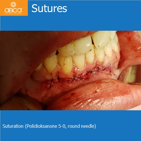
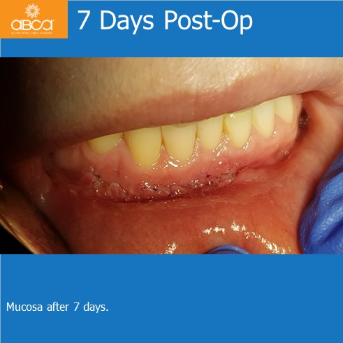
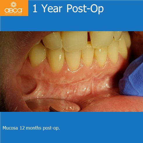
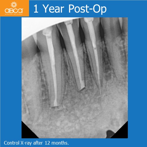
Good healing of the wound was observed. The sutures were removed after 7 days. Moreover, after 12 months no recurrence or secondary inflammatory symptoms were observed.