Bond Apatite - Bone Graft Cement

Bond Apatite - Bone Graft Cement
 
Another novel product of Augma Biomaterials Ltd. is a combination of 3D BondTM with a formula of hydroxyapatite granules. This is a cement-based, osteoconductive composite, synthetic bone substitute that is used for bone reconstruction in a range of dental applications and is intended for filling, augmenting and reconstructing a broad range of defects in the maxillofacial bones.
Free Shipping Get a Free Visa Gift Card
Free Training Kit with Purchase
3D Bond+ Bone Graft Cement (.5cc Syringe) Each  [ AUGMA021-3D-ND-050 ]
Price: $86.70
Quantity:
  
Bond Apatite Bone Graft Cement, (1cc Syringe) Each  [ AUGMA001-HABBPACK1 ]
Price: $135.00
Quantity:
  
Bond Apatite Bone Graft Cement, 1cc (BUY 6 GET 2 FREE) ($96.00 Per Syringe) + Get a Free $50 Visa Gift Card  [ AUGMAPROMO-HABBPACK1 ]
Price: $810.00
Quantity:
  
Bond Apatite Bone Graft Cement, 1cc (BUY 12 GET 5 FREE) ($90.35 Per Syringe) + Get a Free $100 Visa Gift Card  [ AUGMAPROMO-HABBPACK2 ]
Price: $1,620.00
Quantity:
  
Description

Augma Bond Apatite Bone Graft Cement

The fastest, easiest and most efficient bone augmentation material the market has ever seen.

PLACE » PRESS » CLOSE

Another novel product of Augma Biomaterials Ltd. is a combination of 3D BondTM with a formula of hydroxyapatite granules. This is a cement-based, osteoconductive composite, synthetic bone substitute that is used for bone reconstruction in a range of dental applications and is intended for filling, augmenting and reconstructing a broad range of defects in the maxillofacial bones.

Bond Apatite® is composed of 2 matrices which have different absorption coefficients and characteristics.The first matrix is biphasic calcium sulfate (3D BondTM ) which is absorbed and replaced completely. The second matrix is a formula of hydroxyapatite granules which serves as longer range space maintainer. The product guarantees reduced treatment time and convenient manipulation for the clinician, thanks to the product’s unique nature and the specially designed syringe.

  • Bond Apatite® is delivered in a dual-chamber, pre filled syringe,
  • Less than 1 min is required for Graft placement and stabilisation
  • Improved outcomes
  • Shortens Healing Time
  • Membrane use is not essential
  • Significant reduction in overall cost
  • Bond Apatite® is FDA cleared and CE approved


Augma Bone Cement Step-by-step Protocols

Bond Apatite® protocol for socket grafting with flap reflection

Bond Apatite® protocol for socket grafting without flap reflection

3D Bond™ protocol for socket grafting with flap reflection

3D Bond™ protocol for socket without lifting a flap


Recommended Protocols

Socket grafting with 4 bony walls (Can be two ways)

Option 1: without flap reflection


  1. No need to raise a flap
  2. Extract the tooth and prepare the socket for grafting.
  3. Eject the cement into the socket.
  4. Press firmly over the cement for 3 seconds using dry sterile gauze and finger pressure.
    Do not use an instrument to push and compact the cement into the bottom of the socket. (If the interdental space is too narrow to accommodate direct finger pressure on the sterile gauze, then a mirror handle or similar instrument can be applied on top of the gauze).
  5. Protect the cement by covering it with a collagen sponge and secure the sponge in place to the surrounding soft tissue by an initial suture thereafter with a cross stitch above. During the initial stage of healing, the cement should not be left exposed.

Option 2: with flap reflection


Use the same protocol as suggested below for sockets with missing buccal plate.

  1. Before Flap reflection perform short mesial oblique vertical incision (up to 2 mm into the mobile mucosa).
  2. Raise full thickness flap, minimally as needed to expose the entire defect - (Do not preform any manipulation to get tension free flap. No horizontal dissection release cuts , and no brushing. the flap should be with tension during closure and not tension free).
  3. Extract the tooth and prepare the site for grafting
  4. Cement application
    • eject the cement into the site
    • place dry sterile gauze and press firmly for 3 seconds on the buccal and occlusal aspects.
  5. Reposition the flap for maximal closure by stretching it directly above the cement (exposure of 2-3 mm is fine, but no more than that).


Defects with no bony wall frame (Lateral augmentation, ridge widening)


  1. Raise a flap
    • The flap should be minimally reflected in order to expose the entire grafted site. (The vertical cuts should be 2-3 mm into the mobile mucosa) Do not perform any horizontal periosteal dissection for release.
  2. Prepare the site for grafting
  3. Cement application
    • Apply the cement and press firmly for 3 seconds to adapt to the defect using sterile dry gauze.
    • If needed, apply additional layer to obtain desired volume (slightly overfill).
    • Press firmly with the dry sterile gauze for 3 seconds after each layer.
  4. Flap Closure
    • Reposition the flap by stretching it directly above the cement for maximal closure (up to 2-3 mm of graft exposure is fine but not more than that).


Sinus Lift - Lateral window approach protocol


  1. Activate the syringe and wait 1 minute before application.
  2. Eject the cement into the sinus cavity through the sinus lateral window until 2/3 of the sinus is filled (During cement dispersion in the sinus cavity, if needed tap gently above the material with a sterile dry gauze to absorb excess of fluid and blood).
  3. For filling the last 1/3 and closing the sinus window. After activation of the cement (Do not wait 1 minute, eject it immediately into the site, place sterile dry gauze ,press firmly for 3 seconds ,and close the flap .


Sinus Lift - Intra crestal approach protocol


  1. Activate the syringe.
  2. After activation, eject the material into a dish and let it set for 3 minutes.
  3. Use the syringe as a carrier (Any other bone carriers can be used as well).


Biphasic CS bone cement radiographic appearance

** Due to the replacement of the cement into the patients own bone, the Radiographic appearance will vary during the healing period.

  • During graft placement - Radiopaque
  • 2-3 weeks post op - Radiolucent
  • 12 weeks post op - Radiopaque

Biphasic CS technology the foundation for complete bone regeneration

3D Bond™ Bond Apatite®
Socket preservation X X
Simultaneous augmentation of bone defects around Implants X
Bone augmentation in periodontal defects X
Lateral augmentation, ridge expansion, and ridge preservation X
Sinus Lift- Lateral window approach X
Sinus Lift Intra crestal approach X X
Dehisence, fenestrations around teeth and/or Implants X

Table 1: Appropriate indications for 3D Bond™ and Bond Apatite®

* Only recommended in relatively small sockets such as incisives and premolars.


Augma Bond Apatite Publications


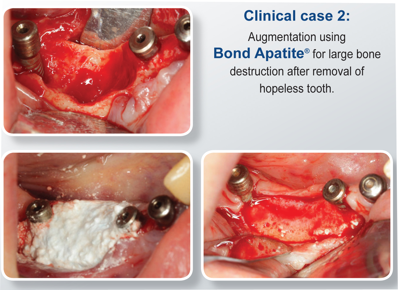
Clinical Cases using Augma Bond Apatite

The Bone Cement Podcast

The Bone Cement Podcast
     

Additional Augma Bond Apatite Resources


Additional Augma Bond Apatite Videos and Images


Bond Apatite Bone Graft Cement - Clinical Case
Bond_Apatite_Augma_Biomaterials

Bone_Graft_Cement_New_Line_Medical

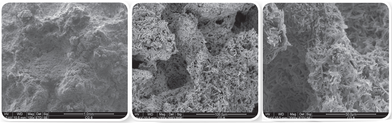
Bond Apatite Internal porosity structure divided into macro porous in different magnifications (SEM images)