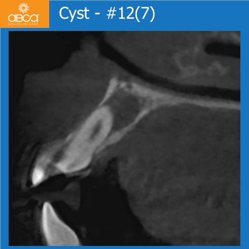
Small Cysts #12 (7) & #22 (10)
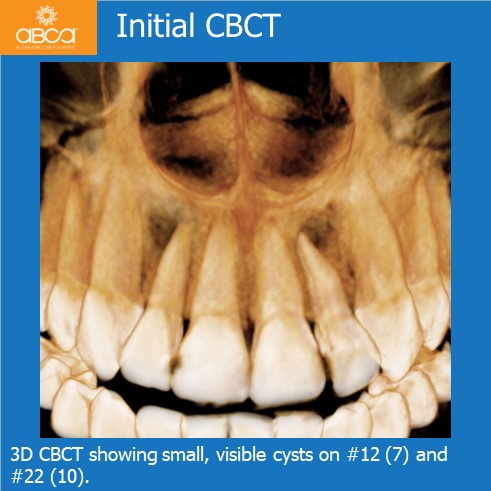
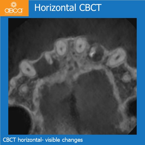
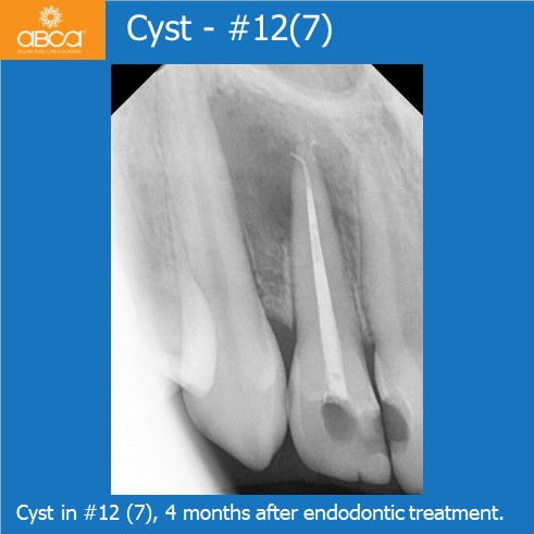
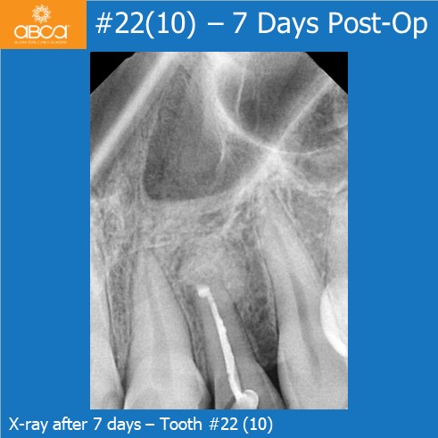
The patient is a healthy, 20 year old female. Two cysts are discovered after she experienced strong pain in area of tooth #12 (7). The cyst is not resolved after 4 months of endodontic treatment. She has occasional pain and discomfort during this period.
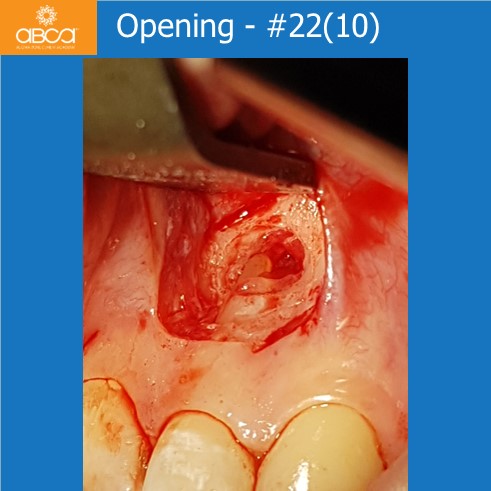
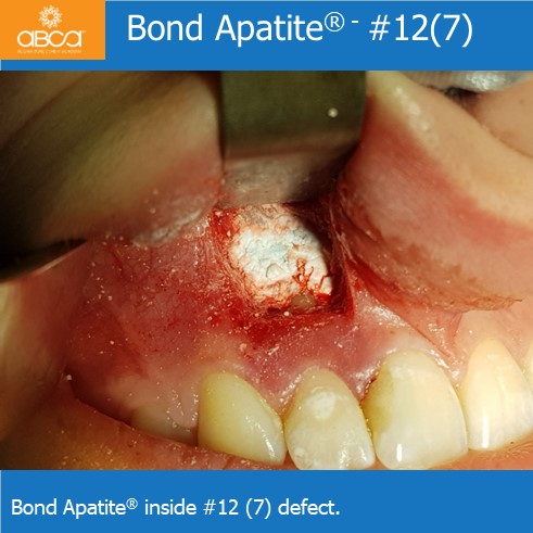
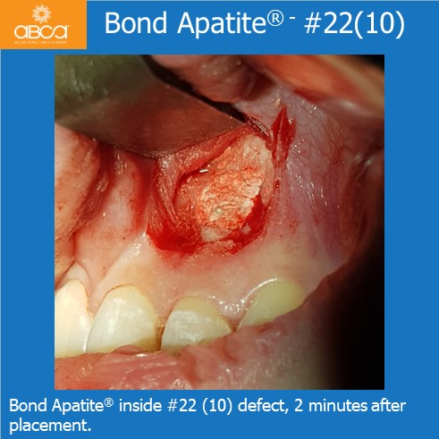
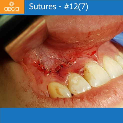
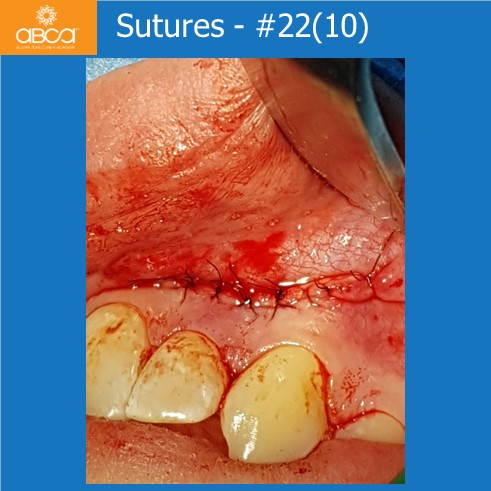
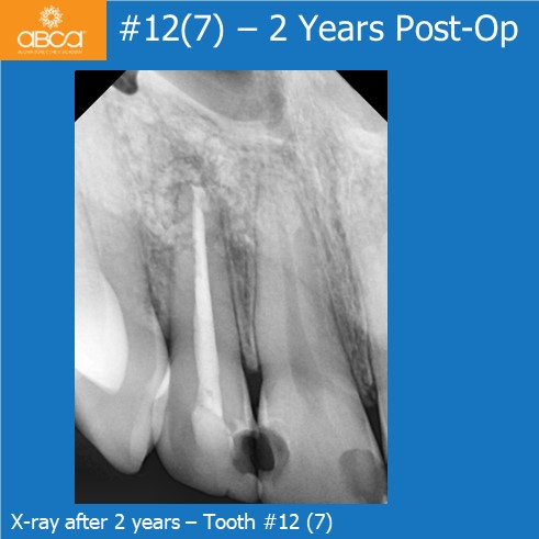
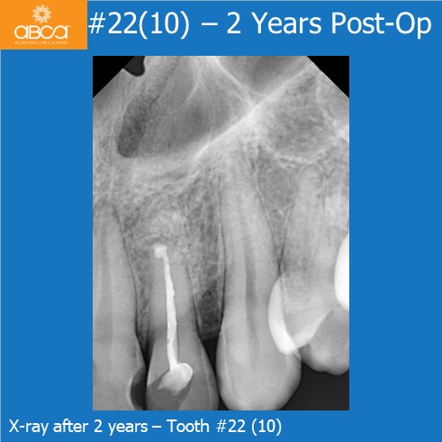
Root resection was performed on teeth #12 (7) and #22 (10). At this same time, cyst enucleation with histopathological examination confirmed radicular cysts. The bone defect was filled with Bond Apatite® (1 cc). More Bond Apatite® was used in #12 (7) and #22 (10).
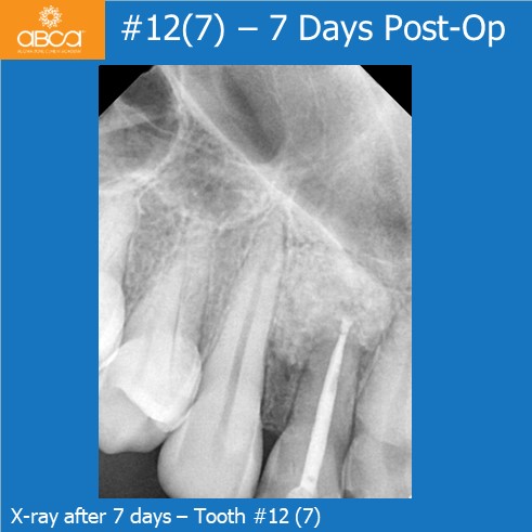
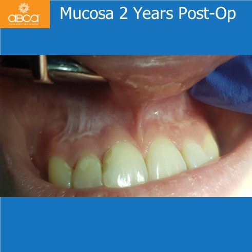
Two year post-op follow up showed good remodeling of the bone, and healing was observed.
Dr. Damian Dudek
DMD, PhD, Poland